


 Foundational characteristics of cancer include proliferation, angiogenesis, migration, evasion of apoptosis, and cellular immortality. Find key markers for these cellular processes and antibodies to detect them.
 The SUMOplot™ Analysis Program predicts and scores sumoylation sites in your protein. SUMOylation is a post-translational modification involved in various cellular processes, such as nuclear-cytosolic transport, transcriptional regulation, apoptosis, protein stability, response to stress, and progression through the cell cycle.
 The Autophagy Receptor Motif Plotter predicts and scores autophagy receptor binding sites in your protein. Identifying proteins connected to this pathway is critical to understanding the role of autophagy in physiological as well as pathological processes such as development, differentiation, neurodegenerative diseases, stress, infection, and cancer.
VAMP7 Antibody
- SPECIFICATION
- CITATIONS
- PROTOCOLS
- BACKGROUND

Application
| WB, IF, E |
|---|---|
| Primary Accession | P51809 |
| Other Accession | NP_001172112, 6845 |
| Reactivity | Human, Mouse, Rat |
| Host | Rabbit |
| Clonality | Polyclonal |
| Isotype | IgG |
| Calculated MW | 24935 Da |
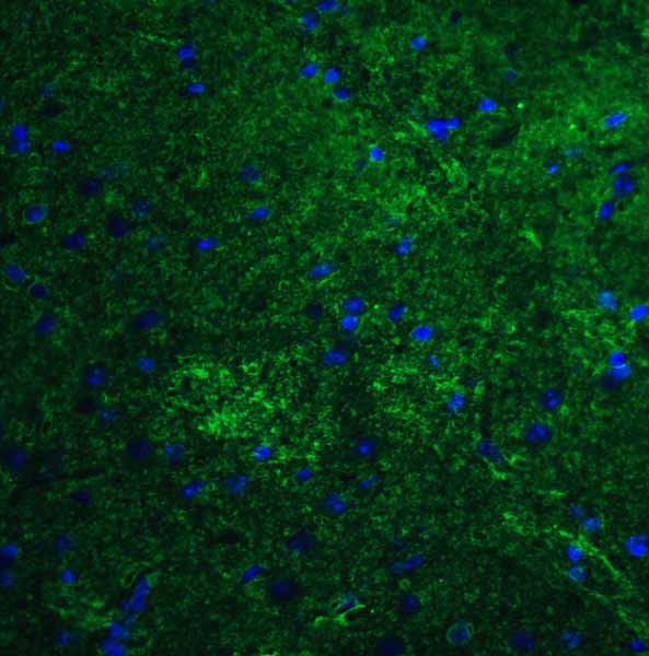
| Application Notes | VAMP7 antibody can be used for detection of VAMP7 by Western blot at 0.5 - 1 μg/mL. For immunofluorescence start at 20 μg/mL. |
| Gene ID | 6845 |
|---|---|
| Target/Specificity | VAMP7 antibody was raised against an 18 amino acid synthetic peptide near the amino terminus of human VAMP7. The immunogen is located within amino acids 20 - 70 of VAMP7. |
| Reconstitution & Storage | VAMP7 antibody can be stored at 4℃ for three months and -20℃, stable for up to one year. As with all antibodies care should be taken to avoid repeated freeze thaw cycles. Antibodies should not be exposed to prolonged high temperatures. |
| Precautions | VAMP7 Antibody is for research use only and not for use in diagnostic or therapeutic procedures. |
| Name | VAMP7 |
|---|---|
| Synonyms | SYBL1 |
| Function | Involved in the targeting and/or fusion of transport vesicles to their target membrane during transport of proteins from the early endosome to the lysosome. Required for heterotypic fusion of late endosomes with lysosomes and homotypic lysosomal fusion. Required for calcium regulated lysosomal exocytosis. Involved in the export of chylomicrons from the endoplasmic reticulum to the cis Golgi. Required for exocytosis of mediators during eosinophil and neutrophil degranulation, and target cell killing by natural killer cells. Required for focal exocytosis of late endocytic vesicles during phagosome formation. |
| Cellular Location | Cytoplasmic vesicle, secretory vesicle membrane; Single-pass type IV membrane protein Golgi apparatus, trans-Golgi network membrane; Single- pass type IV membrane protein. Late endosome membrane; Single-pass type IV membrane protein Lysosome membrane; Single-pass type IV membrane protein. Endoplasmic reticulum membrane; Single-pass type IV membrane protein. Cytoplasmic vesicle, phagosome membrane; Single-pass type IV membrane protein. Synapse, synaptosome. Note=In immature neurons expression is localized in vesicular structures in axons and dendrites while in mature neurons it is localized to the somatodendritic region Colocalizes with LAMP1 in kidney cells. Localization to the endoplasmic reticulum membrane was observed in the intestine but not in liver or kidney (By similarity). |
| Tissue Location | Detected in all tissues tested. |

Thousands of laboratories across the world have published research that depended on the performance of antibodies from Abcepta to advance their research. Check out links to articles that cite our products in major peer-reviewed journals, organized by research category.
info@abcepta.com, and receive a free "I Love Antibodies" mug.

Provided below are standard protocols that you may find useful for product applications.
Background
VAMP7 Antibody: VAMP7 is a member of the soluble N-ethylmaleimide-sensitive factor attachment protein receptor (SNARE) family, localizing to late endosomes and lysosomes. VAMP7 is thought to mediate the fusion of endosomes to their target lysosomes as well as other exocytosis events during phagocytosis and neuritogenesis. VAMP7 interacts with the VPS9 ankyrin repeat protein VARP, a protein that localizes to early endosomes and thought to regulate endosome dynamics. Together with CD82, VAMP7 can modulate the signaling of EGFR by regulating its endocytosis from the plasma membrane.
References
Galli T, Zahraoui A, Vaidyanathan VV, et al. A novel tetanus neurotoxin-insensitive vesicle-associated membrane protein in SNARE complexes of the apical plasma membrane of epithelial cells. Mol. Biol. Cell 1998; 9:1437-48.
Wang Y and Tang BL. SNAREs in neurons - beyond synaptic vesicle exocytosis. Mol. Membr. Biol. 2006; 23:377-84.
Advani RJ, Yang B, Prekeris R, et al. VAMP-7 mediates vesicular transport from endosomes to lysosomes. J. Cell Biol. 1999; 146:765-75.
Burgo A, Sotirakis E, Simmler MC, et al. Role of Varp, a Rab21 exchange factor and TI-VAMP/VAMP7 partner in neurite growth. EMBO Rep. 2009; 10:1117-24.
If you have used an Abcepta product and would like to share how it has performed, please click on the "Submit Review" button and provide the requested information. Our staff will examine and post your review and contact you if needed.
If you have any additional inquiries please email technical services at tech@abcepta.com.
Ordering Information
Other Products
Shipping Information
